Medical Imaging

Speckle contrast optical spectroscopy (SCOS) is an optical technique capable of measuring human cerebral blood flow and brain function non-invasively. Its tomographic extension, speckle contrast optical tomography (SCOT), can provide blood flow variation maps with measurements using overlapping source-detector channel pairs. Linearity is often assumed in most image reconstruction methods, but non-linearity could exist in the relations between measured signals and blood flow variations.
- Categories:
 7 Views
This 3DTeethSegX dataset is a benchmark dataset specifically designed for tooth point cloud completion and segmentation tasks. Built upon the publicly available 3DTeethSeg 2022 MICCAI Challenge dataset, it comprises 1,494 pairs of tooth point clouds and their corresponding tooth images from 38 patients. Each pair includes a partial point cloud (2,048 points) and a complete point cloud (16,384 points).
- Categories:
 32 Views
Brain tumors are one of the most common diseases threatening human health. Early detection and precise segmentation are of great significance for clinical diagnosis and treatment. This paper presents a Learnable Wavelet Transform and Attention Mechanism network(LWTA-Net2D), based on 2D Convolutional Neural Networks (CNN), integrating Learnable Discrete Wavelet Transform (LDWT), combination of Monte Carlo Attention (MCattn) and Monte Carlo Bottleneck Layer (MCBottleneck), and a U-Net-based encoder-decoder architecture.
- Categories:
 16 Views
To enhance the application of radiotherapy planning, we developed a novel head and neck imaging dataset, HND, comprising simulation X-ray computed tomography (CT) images from 486 patients with head and neck cancers who underwent intensity-modulated radiotherapy (IMRT) between February 2019 and June 2024 (Table 1).
- Categories:
 34 Views
This dataset is designed for research on 2D Multi-frequency Electrical Impedance Tomography (mfEIT). It includes:
- Categories:
 68 Views
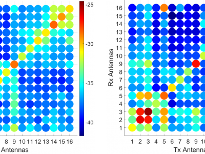
Spread spectrum time domain reflectometry (SSTDR) is proposed to replace the VNA or UWB pulsed systems and switches in a microwave imaging system. These tests evaluate an SSTDR system (Keysight N7081A) from 2-4 GHz. 16 ultrawideband (UWB) antennas were placed in contact with the breast phantom. The McGill breast phantom is a hemispherical carbon-based phantom with the electrical properties of fat. A cylindrical hole allows for the insertion of a plug with fat properties or fat+tumor properties. These were both measured and provided in the attached data set.
- Categories:
 277 Views
The database compiled for this study is a comprehensive and meticulously curated repository designed to evaluate the efficacy of anti-VEGF therapy in patients with Diabetic Macular Edema (DME). It includes clinical and imaging data from 193 diabetic patients, aged 18-70 years, who participated in a single-center, randomized, parallelgroup, double-masked clinical trial. The database encompasses detailed demographic and clinical information, such as age, gender, medical history, duration of diabetes, and baseline measurements like blood pressure and intraocular pressure.
- Categories:
 40 Views


