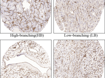
Medical Imaging
This cell images dataset is collected using an ultrafast imaging system known as asymmetric-detection time-stretch optical microscopy (ATOM) for training and evaluation. This novel imaging approach can achieve label-free and high-contrast flow imaging with good cellular resolution images at a very high speed. Each acquired image belongs to one of the four classes: THP1, MCF7, MB231 and PBMC.
- Categories:
Recent advances in scalp electroencephalography (EEG) as a neuroimaging tool have now allowed researchers to overcome technical challenges and movement restrictions typical in traditional neuroimaging studies. Fortunately, recent mobile EEG devices have enabled studies involving cognition and motor control in natural environments that require mobility, such as during art perception and production in a museum setting, and during locomotion tasks.
- Categories:
Ovarian cancer is among the top health issues faced by women everywhere in the world . Ovarian tumours have a wide range of possible causes. Detecting and tracking down these cancers in their early stages is difficult which adds to the difficulty of treatment. In most cases, a woman finds out she has ovarian cancer after it has already spread. In addition, as technology in the field of artificial intelligence advances, detection can be done at an earlier level. Having this data will assist the gynaecologist in treating these tumours as soon as possible.
- Categories:
This dataset consists of both non-retinal detachment and rhegmatogenous retinal detachment fundus images. The fundus images were collected from the four eye hospital in the country (namely India) such as Silchar medical college and hospital (Assam), Aravind eye hospital (Tamil Nadu), LV prasad eye hospital (Hyderabad), and Medanta- The medicity (Gurugram). A total of 1693 images have been collected from these hospitals of which 1017 fundus images belonged to retinal detachments and the rest 676 were non-retinal detachments.
- Categories:

This is the supplemental documents which include the latex file and figure files for the manuscript titled "Accelerating Magnetic Resonance T1ρ Mapping Using Simultaneously Spatial Patch-based and Parametric Group-based Low-rank Tensors (SMART) ".
- Categories:
Fabdepth HMI is designed for hand gesture detection for Human Machine Interaction. It contains total of 8 gestures performed by 150 different individuals. These individuals range from toddlers to senior citizens which adds diversity in this dataset. These gestures are available in 3 different formats namely resized, foreground=-background separated and depth estimated images. Additional aspect is added in terms of video format of 150 samples. Researchers may choose their combination of data modalities based on their application.
- Categories:

We provide the abstract from the paper below:
- Categories:

The datset inculed the BrainWeb data which consists of T1-weighted (T1w), T2-weighted (T2w), and proton density-weighted (PDw) normal brain noise-free MR images (the size is with resolution), two real T1w MR brain datasets (OAS30040 and OAS30072) from the Open Access Series of imaging Studies (OASIS) database,and the synthetic DW-MRI dataset
- Categories:

Retinal Fundus Multi-disease Image Dataset (RFMiD 2.0) is an auxiliary dataset to our previously published dataset. RFMiD 2.0 is a more challenging dataset to research society to develop the computer-based disease diagnosis system. Diabetic Retinopathy, cataracts, and refractive error in the eye are leading disease which causes permanent vision loss more frequently. Therefore, developing an AI-based model to classify these diseases is useful for ophthalmologists. This dataset consists of 860 images of frequently and rarely observed 51 diseases.
- Categories:
This is just a preliminary collation of the relevant TCGA datasets collated and used in our methodology. We will continue to upload the full dataset later for your reference and use. We hope to make a small contribution to the study of automatic 3D MRI classification of gliomas and the problem of domain adaptation on medical images.
- Categories: