Dental OPG XRAY Dataset
- Citation Author(s):
-
Rubaba Binte Rahman (American International University-Bangladesh)Sharia Arfin Tanim
 (American International University-Bangladesh)
Nazia Alfaz (American International University-Bangladesh)Tahmid Enam Shrestha (American International University-Bangladesh)M Saef Ullah Miah (American International University-Bangladesh)Firoz Mridha (American International University-Bangladesh)
(American International University-Bangladesh)
Nazia Alfaz (American International University-Bangladesh)Tahmid Enam Shrestha (American International University-Bangladesh)M Saef Ullah Miah (American International University-Bangladesh)Firoz Mridha (American International University-Bangladesh) - Submitted by:
- Sharia Tanim
- Last updated:
- DOI:
- 10.21227/tzrj-ws66
- Data Format:
- Research Article Link:
 682 views
- Categories:
- Keywords:
Abstract
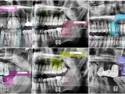
This article presents a dental dataset for the improvement of research on deep learning-based detection and classification of dental diseases. The dataset is consisted of 232 panoramic dental radiographs, categorized into six major classes: healthy teeth, caries, impacted teeth, infections, fractured teeth, and broken-down crowns/roots (BDC/BDR). The images were collected from three renowned private clinics in Dhaka, Bangladesh, with the help of an experienced dental practitioner who ensured the confidentiality of patients and high-quality data acquisition using a 64-megapixel Android phone camera. To enhance the value of the dataset for machine and deep learning applications, we applied Contrast-Limited Adaptive Histogram Equalization (CLAHE) for image enhancement and augmented the data. The images were annotated using the CVAT tool and reviewed by dental experts. This benchmark dataset is publicly available and provides a valuable resource for researchers in artificial intelligence, computer science, and dental informatics to promote interdisciplinary collaboration and the development of advanced algorithms for dental disease detection.
Instructions:
Description
This dataset includes dental OPG X-rays collected from three different dental clinics. This dataset can be used for tasks like object detection, image analysis, disease classification, and segmentation. It has two folders: the object detection dataset folder and the classification dataset folder. The object detection folder contains 232 original and 604 augmented images and labels. The classification folder contains six distinct files for each class. The images are in JPG format, and the labels are in JSON format. The augmented data is split into training, validation, and testing sets in an 80:10:10 ratio. Dataset collection: • Source: Prescription Point Ltd, Lab Aid Specialized Hospital, Ibn Sina Diagnostic and Imaging Center. • Capture Method: Using android phone camera. • Anonymization: All data were rigorously anonymized to maintain confidentiality and privacy. • Informed Consent: All patients provided their consent in accordance with the dental ethical principles. Dataset composition: • Total Participants: 232 Male and female patients aged 10 years or older. Variables: • Healthy Teeth: 223 • Caries: 119 • Impacted Teeth: 87 • Broken Down Crown/ Root: 52 • Infection: 23 • Fractured Teeth: 13


thank you