stroke

This data set contains:


-88 patients


-the noncontrast computed tomography (NCCT) and computed tomography angiography (CTA) performed before thrombectomy.


-the VOI of blood clot for NCCT and CTA.


For each patient NCCT data is marked "2" and CTA is marked "1".

Categories:
644 Views

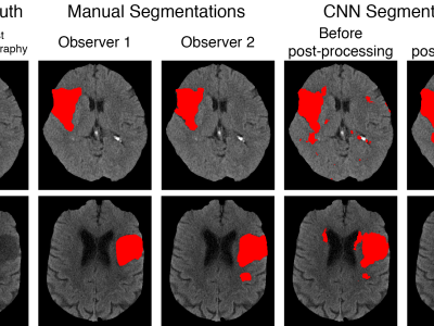
This dataset contains the trained model that accompanies the publication of the same name:

 Anup Tuladhar*, Serena Schimert*, Deepthi Rajashekar, Helge C. Kniep, Jens Fiehler, Nils D. Forkert, "Automatic Segmentation of Stroke Lesions in Non-Contrast Computed Tomography Datasets With Convolutional Neural Networks," in IEEE Access, vol. 8, pp. 94871-94879, 2020, doi:10.1109/ACCESS.2020.2995632. *: Co-first authors


Categories:
2791 Views

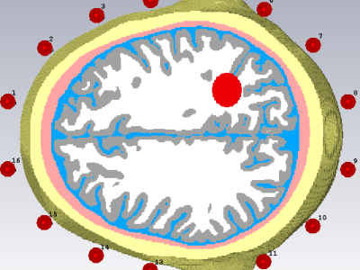
The proposed signals are used  for electromagnetic-based stroke classification.  Six realistic head phantom computed from MRI scans, is surrounded by an antenna array of 16 dipole antennas distributed uniformly around the head. These antennas are deployed in a fixed circular array around the head, at a distance of approximately 2-3 mm from the head. A Gaussian pulse covering the bandwidth from 0:7 to 2 GHz is emitted from each of the antennas, sequentially, while all of the antennas capture the scattered signals. Since 16 antennas were used, there are a total of 256 channel signals (i.e.

Categories:
3921 Views