microscopy

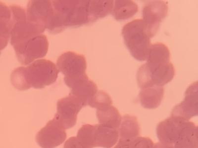
This dataset consists of 462 field of views of Giemsa(dye)-stained and field(dye)-stained thin blood smear images acquired using an iPhone 10 mobile phone with a 12MP camera. The phone was attached to an Olympus microscope with 1000× objective lens. Half of the acquired images are red blood cells with a normal morphology and the other half have a Rouleaux formation morphology.

Categories:
863 Views

This <file> was prepared along with the establishment of the reference intervals for the hematological and biochemical parameters of the juvenile Visayan warty pig (Sus cebifrons negrinus).  The determination of the reference intervals of the juvenile Visayan warty pig was significant, as these reference intervals have never been established for the purpose of assessing the health of this critically endangered species. The file contains six separate sheets featuring the raw and transformed data, as well as the calculated reference interval.

Categories:
409 Views

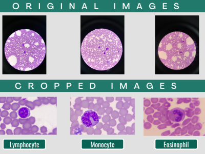
This dataset was prepared to aid in the creation of a machine learning algorithm that would classify the white blood cells in thin blood smears of juvenile Visayan warty pigs. The creation of this dataset was deemed imperative because of the limited availability of blood smear images collected from the critically endangered species on the internet. The dataset contains 3,457 images of various types of white blood cells (JPEG) with accompanying cell type labels (XLSX).

Categories:
3295 Views

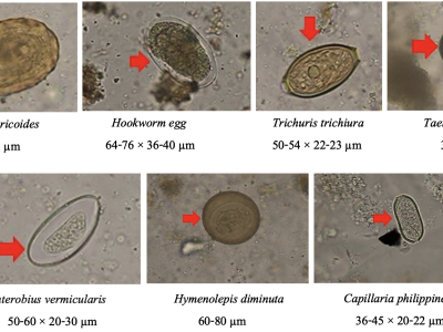
Parasitic infections have been recognised as one of the most significant causes of illnesses by WHO. Most infected persons shed cysts or eggs in their living environment, and unwittingly cause transmission of parasites to other individuals. Diagnosis of intestinal parasites is usually based on direct examination in the laboratory, of which capacity is obviously limited.

Last Updated On: 
Mon, 02/26/2024 - 10:24
Citation Author(s): 
Duangdao Palasuwan, Thanarat H. Chalidabhongse, Korranat Naruenatthanaset, Thananop Kobchaisawat, Kanyarat Boonpeng, Nuntiporn Nunthanasup, Nantheera Anantrasirichai

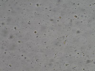
Fecal microscopic data set is a set of fecal microscopic images, which is used in object detection task. The datasets are collected from the Sixth People’s Hospital of Chengdu (Sichuan Province, China). The samples were went flow diluted, stirred and placed, and imaged with a microscopic imaging system. The clearest 5 images were collected for each view of each sample with Tenengrad definition algorithm. The dataset we collected includes 10670 groups of views with 53350 jpg images. The Resolution of images are 1200×1600. There are 4 categories, RBCs, WBCs, Molds, and Pyocytes.

Categories:
666 Views