MRI

This dataset contains MRI data acquired approximately 20 minutes before ("Pre-ablation") and 20-40 minutes after ("Post-ablation") MR-guided focused ultrasound thermal ablations in the muscle tissue (quadriceps) in four (n=4) New Zealand white rabbits. MR images include MR thermometry acquired with the proton resonance frenquency method, ADC maps (b=0,400), T2-weighted, and pre- and post-contrast enhanced T1-weighted images. All MRI acquisitions were 3D except for the ADC acquisition, which was 2D multi-slice.

Categories:
149 Views

Use of medical devices in the magnetic resonance environment is regulated by standards that include the ASTM-F2213 magnetically induced torque. This standard prescribes five tests. However, none can be directly applied to measure very low torques of slender lightweight devices such as needles. Methods: We present a variant of an ASTM torsional spring method that makes a “spring” of 2 strings that suspend the needle by its ends. The magnetically induced torque on the needle causes it to rotate. The strings tilt and lift the needle.

Categories:
273 Views

Neuroimaging methods play an important role in presurgical examinations and localization of epileptogenic lesion. Magnetic resonance imaging (MRI) is a neuroimaging technique that is essential to detect structurally abnormal tissue and thus delineate the epileptogenic lesion. Magnetic resonance imaging (MRI) provides structural data and can reveal underlying epileptogenic lesions (T1, T2, FLAIR).

Categories:
500 Views

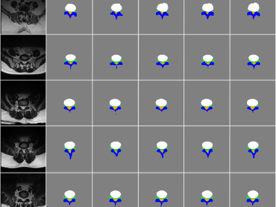
Research data associated with paper: A Semantic Segmentation Model for Lumbar MRI Images using Divergence Loss, comprising the python code, a trained model and empirical results. 

Categories:
366 Views

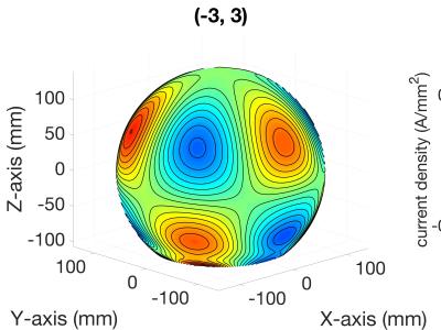
<address>Designed Shim Coil Layouts</address><address>Use Visualize_Coils.m with Matlab 2018a can visualize SH coils.</address>

Categories:
112 Views


Example axial and coronal phase maps and post-treatment MRI from 68 thalamotomies in essential tremor patients and four pallidotomies in Parkinson's disease patients. From the manuscript "Using phase data from MR temperature imaging to visualize anatomy during MRI-guided focused ultrasound neurosurgery" published in 2020 in IEEE Trans. Med. Imaging.


Normal
0

false
false
false

EN-US
X-NONE
X-NONE

Categories:
387 Views

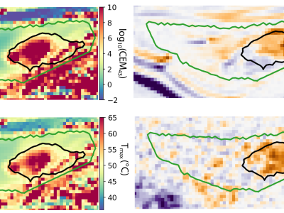
The following pages show axial T2-weighted MRI obtained at 24 hours and at 3-15 months after MRgFUS. The images shown here were registered to the same reference frame that was used in the thermal simulations; every third image is shown. To segment the bone marrow lesions, the registered images were toggled back and forth between the two time points to detect obvious changes. The lesion segmentations were completed before the acoustic and thermal simulations were performed. They were originally done on the native T2-weighted images acquired at 3-15 months after FUS.

Categories:
257 Views

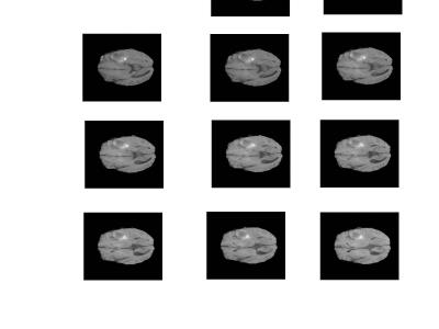
BraTS has always been focusing on the evaluation of state-of-the-art methods for the segmentation of brain tumors in multimodal magnetic resonance imaging (MRI) scans. BraTS 2019 utilizes multi-institutional pre-operative MRI scans and focuses on the segmentation of intrinsically heterogeneous (in appearance, shape, and histology) brain tumors, namely gliomas. Furthemore, to pinpoint the clinical relevance of this segmentation task, BraTS’19 also focuses on the prediction of patient overall survival, via integrative analyses of radiomic features and machine learning algorithms.

Last Updated On: 
Fri, 02/28/2020 - 06:31