Unpaired MR-CT Brain Dataset for Unsupervised Image Translation
- Citation Author(s):
- Submitted by:
- Omar Al-Kadi
- Last updated:
- DOI:
- 10.21227/c9yx-x936
- Data Format:
- Links:
 1785 views
- Categories:
- Keywords:
Abstract
The Magnetic Resonance – Computed Tomography (MR-CT) Jordan University Hospital (JUH) dataset has been collected after receiving Institutional Review Board (IRB) approval of the hospital and consent forms have been obtained from all patients. All procedures followed are consistent with the ethics of handling patients’ data.
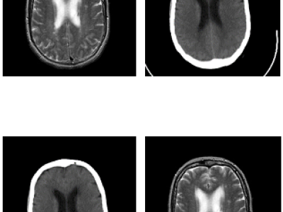
The dataset consists of unpaired brain CT and MR images of 20 patients scanned for radiotherapy treatment planning for brain tumors. The dataset contains T2-MR and CT images for patients aged between 26-71 years with mean-std equal to 47-14.07. The MR images were acquired with a 5.00mm T Siemens Verio 3T using a T2-weighted without contrast agent, 3 Fat sat pulses (FS), 2500-4000 TR, 20-30 TE, and 90/180 flip angle. The CT images were acquired with Siemens Somatom scanner with 2.46mGY.cm dose length, 130KV voltage, 113-327 mAs tube current, topogram acquisition protocol, 64 dual source, one projection, and slice thickness of 7.0mm. Smooth and sharp filters have been applied to CT images. The MR scans have a resolution of 0.7×0.6×5 mm^3, while the CT scans have a resolution of 0.6×0.6×7 mm^3. There are a total of 420 2D axial image slices in the compressed folder (420 MR and 420 CT 2D axial image slices). (Please contact Omar Al-Kadi at o.alkadi[at]ju.edu.jo for compressed file access code)
Instructions:
The Magnetic Resonance – Computed Tomography (MR-CT) Jordan University Hospital (JUH) dataset has been collected after receiving Institutional Review Board (IRB) approval of the hospital and consent forms have been obtained from all patients. All procedures followed are consistent with the ethics of handling patients’ data.
The dataset consists of unpaired brain CT and MR images of 20 patients scanned for radiotherapy treatment planning for brain tumors. The dataset contains T2-MR and CT images for patients aged between 26-71 years with mean-std equal to 47-14.07. The MR images were acquired with a 5.00mm T Siemens Verio 3T using a T2-weighted without contrast agent, 3 Fat sat pulses (FS), 2500-4000 TR, 20-30 TE, and 90/180 flip angle. The CT images were acquired with Siemens Somatom scanner with 2.46mGY.cm dose length, 130KV voltage, 113-327 mAs tube current, topogram acquisition protocol, 64 dual source, one projection, and slice thickness of 7.0mm. Smooth and sharp filters have been applied to CT images. The MR scans have a resolution of 0.7×0.6×5 mm^3, while the CT scans have a resolution of 0.6×0.6×7 mm^3. There are a total of 840 2D axial image slices in the compressed folder (420 MR and 420 CT 2D axial image slices). (Please contact Omar Al-Kadi at o.alkadi[at]ju.edu.jo for compressed file access code)


