Light-field microscopy data
- Citation Author(s):
-
Pingfan Song (Imperial College London)
- Submitted by:
- Pingfan Song
- Last updated:
- DOI:
- 10.21227/864p-p592
- Data Format:
- Research Article Link:
 651 views
- Categories:
- Keywords:
Abstract
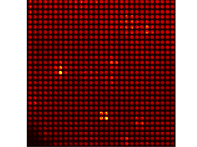
This dataset contains light-field microscopy images and converted sub-aperture images.
The folder with the name "Light-fieldMicroscopeData" contains raw light-field data. The file LFM_Calibrated_frame0-9.tif contains 9 frames of raw light-field microscopy images which has been calibrated. Each frame corresponds to a specific depth. The 9 frames cover a depth range from 0 um to 32 um with step size 4 um. Files with name LFM_Calibrated_frame?.png are the png version for each frame.
The folder with the name "SubapertureImgsArray" contains sub-aperture images converted from raw light-field images.
Instructions:
Please enter the folder with the name "Light-fieldMicroscopeData". Use FIJI or other software for viewing images to examine the raw light-field data "LFM_Calibrated_frame0-9.tif". In each light-field image, we can see an array of small round spots which are the back-aperture of lenslets.
Please enter the folder with the name "SubapertureImgsArray". Use FIJI or other software for viewing images to examine sub-aperture image arrays for different depths with the name "SubapertureImgsArray_frame?.png". Each sub-aperture image in an array is composed of pixels that share the same relative position behind each microlens.


it will be helpful to the research in light field imaging