Datasets
Standard Dataset
Rouleaux Morphology Red Blood Cells
- Citation Author(s):
- Submitted by:
- FATIMA MUHAMMAD
- Last updated:
- Fri, 07/26/2024 - 19:56
- DOI:
- 10.21227/wed3-1619
- Data Format:
- Research Article Link:
- License:
 1017 Views
- Categories:
- Keywords:
Abstract
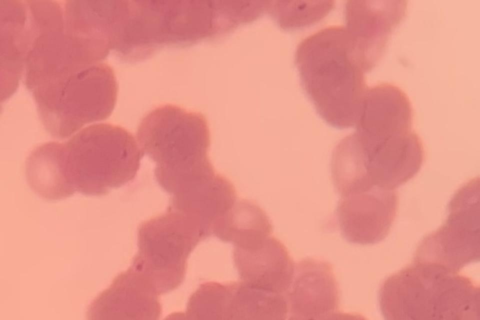
This dataset consists of 462 field of views of Giemsa(dye)-stained and field(dye)-stained thin blood smear images acquired using an iPhone 10 mobile phone with a 12MP camera. The phone was attached to an Olympus microscope with 1000× objective lens. Half of the acquired images are red blood cells with a normal morphology and the other half have a Rouleaux formation morphology. Out of the Rouleaux images, 51 images were acquired from Giemsa-stained slides at Murtala Muhammad Specialist hospital, Kano state, Nigeria while 180 images were acquired from field-stained slides at Isyaka Rabiu paediatrics hospital, Kano state, Nigeria. The 231 normal cell morphology images were acquired from Murtala Muhammad specialist hospital from slides stained with Giemsa. The slides used consists of cells of varying morphology such as crenated cells, microcytes, ovalocytes, normalocytes and sickle cells. The size of the original captured images are in the range of 4032×3024 which were cropped and sliced to 750x750 to ease computational requirements during model training. If the original captured images are required, the author can be contacted.
This dataset consists of two files. A File with images of red blood cells having a normal morphology (Normal red blood cells) and the other file with images of red blood cells with rouleaux formation morphology (Rouleaux Red blood cells). Each file contains equal instances of 3,044 images with a size of 750x750 intended for binary classification.
Dataset Files
- DATA SET.zip (518.84 MB)
- Whole field of View (FOV) images Stage 1 Cropped.zip (382.59 MB)


Comments
Edited