Biomedical and Health Sciences

This repository aims to publish a sEMG database for hand gesture recongnition, which is suitable for intra-session, inter-session, inter-day and inter-subject tests. Six subjects were involved in data collection on ten days, and two sessions a day with the interval of half an hour. In each session, one trial (10 secondes) for each geature was conducted. The electrode sleeve did not reweared between two sessions in a day. The utilised sEMG device was customised by the Intelligent System and Biomedical Robotics Group, which was discussed in [1].
- Categories:
 1211 Views
# ISRMyo-I: A Database for sEMG-based Hand Gesture Recognition
## Introduction
- Categories:
962 ViewsAccess the dataset for images of typical diabetic retinopathy lesions and also normal retinal structures annotated at a pixel level, focused on an Indian population. This dataset provides information on the disease severity of diabetic retinopathy, and diabetic macular edema for each image.
- Categories:
119144 Views
For research purposes, the ECG signals were obtained from the PhysioNet service (http://www.physionet.org) from the MIT-BIH Arrhythmia database. The created database with ECG signals is described below. 1) The ECG signals were from 29 patients: 15 female (age: 23-89) and 14 male (age: 32-89). 2) The ECG signals contained 17 classes: normal sinus rhythm, pacemaker rhythm, and 15 types of cardiac dysfunctions (for each of which at least 10 signal fragments were collected).
- Categories:
10755 Views
The published sEMG database was captured by the Intelligent System and Biomedical Robotics Group at University of Portsmouth, leaded by Prof. Honghai Liu.
Six subjects were volunteered for data capturing, and the sEMG data were captured in ten separate days. We manually separated the whole database into two parts: training dataset (the first 7 days) and testing dataset(the last 3 days). For each subject, two folders exist, one for training and the other for test.
- Categories:
551 Views
TB (Tuberculosis) is a contagious disease which is caused by a bacterium named Mycobacterium Tuberculosis. Screening is done to confirm the presence of TB using different screening techniques available i.e. Chest X-ray, Microscopy, Gene Xpert and Culture etc. Medical image processing is a rapidly growing field of image processing that is used to automate different medical procedures. In this research we have designed two automated systems for the screening of TB patients. A sample of 50 images for microscopy slides and chest X-ray radiographs were taken.
- Categories:
572 Views
TB (Tuberculosis) is a contagious disease which is caused by a bacterium named Mycobacterium Tuberculosis. Screening is done to confirm the presence of TB using different screening techniques available i.e. Chest X-ray, Microscopy, Gene Xpert and Culture etc. Medical image processing is a rapidly growing field of image processing that is used to automate different medical procedures. In this research we have designed two automated systems for the screening of TB patients. A sample of 50 images for microscopy slides and chest X-ray radiographs were taken.
- Categories:
832 Views
72
Normal
0
false
false
false
EN-US
X-NONE
X-NONE
/* Style Definitions */
table.MsoNormalTable
{mso-style-name:"Table Normal";
mso-tstyle-rowband-size:0;
mso-tstyle-colband-size:0;
mso-style-noshow:yes;
mso-style-priority:99;
mso-style-parent:"";
mso-padding-alt:0in 5.4pt 0in 5.4pt;
mso-para-margin:0in;
mso-para-margin-bottom:.0001pt;
mso-pagination:widow-orphan;
font-size:10.0pt;
font-family:"Times New Roman",serif;}
- Categories:
183 Views
This cell images dataset is collected using an ultrafast imaging system known as asymmetric-detection time-stretch optical microscopy (ATOM) for training and evaluation. This novel imaging approach can achieve label-free and high-contrast flow imaging with good cellular resolution images at a very high speed. Each acquired image belongs to one of the four classes: THP1, MCF7, MB231 and PBMC.
- Categories:
4238 Views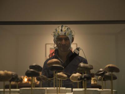
Recent advances in scalp electroencephalography (EEG) as a neuroimaging tool have now allowed researchers to overcome technical challenges and movement restrictions typical in traditional neuroimaging studies. Fortunately, recent mobile EEG devices have enabled studies involving cognition and motor control in natural environments that require mobility, such as during art perception and production in a museum setting, and during locomotion tasks.
- Categories:
4052 Views