Automated Identification of Stereoelectroencephalography Contacts
- Citation Author(s):
- Submitted by:
- Radek Janca
- Last updated:
- DOI:
- 10.21227/5fzx-sv76
- Data Format:
- Research Article Link:
 385 views
- Categories:
- Keywords:
Abstract
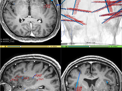
Objective: Stereoelectroencephalography (SEEG) is an established invasive diagnostic technique for use in patients with drug-resistant focal epilepsy evaluated before resective epilepsy surgery. The factors that influence the accuracy of electrode implantation are not fully understood. Adequate accuracy prevents the risk of major surgery complications. Precise knowledge of the anatomical positions of individual electrode contacts is crucial for the interpretation of SEEG recordings and subsequent surgery. Methods: We developed an image processing pipeline to localize implanted electrodes and detect individual contact positions using computed tomography (CT), as a substitute for time-consuming manual labeling. The algorithm automates measurement of parameters of the electrodes implanted in the skull (bone thickness, implantation angle and depth) for use in modeling of predictive factors that influence implantation accuracy. Results: The automated detector localized all contacts with better accuracy than manual labeling (p < 0.001).
The dataset contains MATLAB tool using SPM12 toolbox to detect SEEG electrodes which label all contacts on them. The graphical tutorial is added with sample of CT and MRI images, and stereotactic Luminant template of 61 adults.
Instructions:
User manual and instruction for toolbox use,
Check the latest version at:
https://github.com/EpiReC-ISARG/SEEG-contact-detection-and-skull-measurement


