ADAM: Automatic Detection challenge on Age-related Macular degeneration
- Citation Author(s):
-
Huazhu Fu (Inception Institute of Artificial Intelligence)Fei Li (Zhongshan Ophthalmic Center, Sun Yat-sen University, China.)José Ignacio OrlandoHrvoje Bogunović (Medical University of Vienna)Xu Sun (Baidu Inc., China.)Jingan Liao (South China University of Technology)Yanwu Xu (Baidu Inc., China.)Shaochong Zhang (Zhongshan Ophthalmic Center, Sun Yat-sen University, China.)Xiulan Zhang (Zhongshan Ophthalmic Center, Sun Yat-sen University, China.)
- Submitted by:
- Huazhu Fu
- Last updated:
- DOI:
- 10.21227/dt4f-rt59
- Links:
 4684 views
- Categories:
- Keywords:
Abstract
ADAM is organized as a half day Challenge, a Satellite Event of the ISBI 2020 conference in Iowa City, Iowa, USA.
The ADAM challenge focuses on the investigation and development of algorithms associated with the diagnosis of Age-related Macular degeneration (AMD) and segmentation of lesions in fundus photos from AMD patients. The goal of the challenge is to evaluate and compare automated algorithms for the detection of AMD on a common dataset of retinal fundus images. We invite the medical image analysis community to participate by developing and testing existing and novel automated fundus classification and segmentation methods.
Instructions:
ADAM: Automatic Detection challenge on Age-related Macular degeneration
Link: https://amd.grand-challenge.org
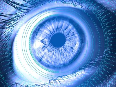
Age-related macular degeneration, abbreviated as AMD, is a degenerative disorder in the macular region. It mainly occurs in people older than 45 years old and its incidence rate is even higher than diabetic retinopathy in the elderly.
The etiology of AMD is not fully understood, which could be related to multiple factors, including genetics, chronic photodestruction effect, and nutritional disorder. AMD is classified into Dry AMD and Wet AMD. Dry AMD (also called nonexudative AMD) is not neovascular. It is characterized by progressive atrophy of retinal pigment epithelium (RPE). In the late stage, drusen and the large area of atrophy could be observed under ophthalmoscopy. Wet AMD (also called neovascular or exudative AMD), is characterized by active neovascularization under RPE, subsequently causing exudation, hemorrhage, and scarring, and will eventually cause irreversible damage to the photoreceptors and rapid vision loss if left untreated.
An early diagnosis of AMD is crucial to treatment and prognosis. Fundus photo is one of the basic examinations. The current dataset is composed of AMD and non-AMD (myopia, normal control, etc.) photos. Typical signs of AMD that can be found in these photos include drusen, exudation, hemorrhage, etc.
The ADAM challenge has 4 tasks:
Task 1: Classification of AMD and non-AMD fundus images.
Task 2: Detection and segmentation of optic disc.
Task 3: Localization of fovea.
Task 4: Detection and Segmentation of lesions from fundus images.


In reply to for academic use by Yalin Zheng
In reply to for academic use by Yalin Zheng
can i use this dataset for learning
In reply to for academic use by Yalin Zheng
Hello, my name is Nayeon Jang. I am a graduate student at Sungkyunkwan Univ. in South Korea, conducting medical AI research in Professor Choo’s lab.
I am currently conducting research on Age-related Macular Degeneration (AMD), and I would like to kindly request access to the ADAM: Automatic Detection Challenge on Age-related Macular Degeneration dataset for academic purposes.
for academic use
I'd like to gain access to the data for academic purposes.
for academic use