Pulmonary lobe segmentation of COVID-19 CT scans
- Citation Author(s):
-
Velmurugan Balasubramanian (Indian institute of technology, Kharagpur)Suhasini Balasubramaniam (Government medical college, Omandurar estate, Chennai)Debdoot Sheet (Indian institute of technology, Kharagpur)Rachana Sathish (Indian institute of technology, Kharagpur)Mahalakshumi Visvanathan (Sri Venkateswara college of engineering, Chennai)
- Submitted by:
- Velmurugan Balasubramanian
- Last updated:
- DOI:
- 10.21227/3qe9-e178
- Data Format:
 1612 views
Abstract
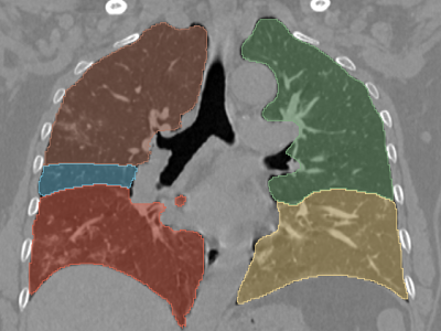
We chose 8 publicly available CT volumes of COVID-19 positive patients which were available from https://doi.org/10.5281/zenodo.3757476 and used 3D slicer to generate volumetric annotations of 512*512 dimension for 5 lung lobes namely right upper lobe, right middle lobe, right lower lobe, left upper lobe and left lower lobe. These annotations are validated by a radiologist with over 15 years of experience.
Instructions:
CT volumes can be downloaded from https://doi.org/10.5281/zenodo.3757476
Volumetric annotations for 5 lobe segments namely right upper lobe, right middle lobe, right lower lobe, left upper lobe and left lower lobe are saved as segments 1 to 5 respectively.
For scans with prefix coronacases_00x their corresponding annotations are uploaded with suffix lobes
The scans and annotations measure 512*512 and are in .nii format