Artificial Intelligence; Dataset; Ultrasound; Machine Learning; Health; Biomedical and Health Sciences; Medical Imaging

A total of 1035 color Doppler US images of heart patients, who were suffering from MR has been collected from the department of cardiology, Swami Rama Himalayan University (SHRU), Dehradun, India. The US images (800 × 600 pixels) used for the analysis of MR were recorded by Philips US machine equipped with multi-frequency transducers of 2-5 MHz range. The images were collected in three different views, i.e., A2C, A4C and PLAX view. 

Categories:

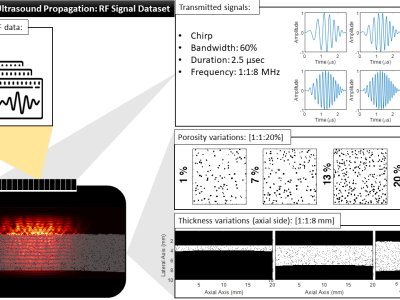
This dataset contains RF (Radio Frequency) signals obtained from simulations, which model ultrasound propagation in cortical bone.

The simulations were designed to provide insights into the behaviour of ultrasound waves in cortical bone tissues, both in intact and pathological conditions. The dataset covers a wide range of parameters, including varying thickness (1-8 mm), porosity (1-20%), and frequency (1-8 MHz), allowing to explore the impact of these factors on ultrasound signal characteristics.


Categories: