Datasets
Standard Dataset
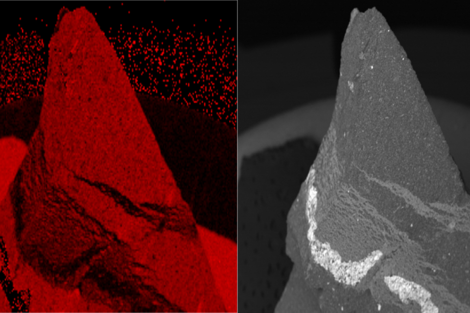
Low-Resolution Multispectral EDS - High-Resolution Panchromatic SEM images for close-range Pansharpening testing.
- Citation Author(s):
- Submitted by:
- Tuomas Sihvonen
- Last updated:
- Fri, 12/22/2023 - 03:36
- DOI:
- 10.21227/zxvc-y179
- Data Format:
- License:
 591 Views
- Categories:
- Keywords:
Abstract
The presented dataset is a supplementary material to the paper [1] and it represents the X-Ray Energy Dispersive (EDS)/ Scanning Electron Microscopy (SEM) images of a shungite-mineral particle. Pansharpening is a procedure for enhancing the spatial resolution of a multispectral image, here the EDS individual bands, with a high-spatial panchromatic image, here the SEM image. Pansharpening techniques are usually tested with remote sensed data, but the procedures have been efficient in close-range MS-PAN pairs as well [3]. The current dataset provides both the MS data in multiple elemental bands and the PAN image associated with the particle angle. All images were captured by a Hitachi SU3500 Scanning Electron Microscope with Thermo Scientific UltraDry SDD EDS, dual detector. The sample is of an uneven nature and presents noise that can be pretreated as in [2], and both the noise-treated and original images are present in the current dataset. The image dimensions are 256 by 192 for the EDS maps and 1024 by 768 for the SEM images, whereas the chemical elements present in the sample are: aluminum (Al), carbon (C), iron (Fe), oxygen (O) and sulfur (S).
[1] T. Sihvonen, Z.-S. Duma and S.-P. Reinikainen, “AB-PLS-DA: Pansharpening tailored for scanning electron microscopy and energy-dispersive X-ray spectrometry multimodal fusion,” Micron, 2024, https://doi.org/10.1016/j.micron.2023.103578
[2] Z.-S. Duma, T. Sihvonen, V. Reinikainen, J. Havukainen, and S.-P. Reinikainen, “Optimizing Energy Dispersive X-Ray Spectroscopy (EDS) Image Fusion to Scanning Electron Microscopy (SEM) Images”, Micron, 2022, https://doi.org/10.1016/j.micron.2022.103361
[3] G. Franchi, J. Angulo, M. Moreaud, and L. Sorbier, “Enhanced EDX images by fusion of multimodal SEM images using pansharpening techniques,” Journal of Microscopy, vol. 269, no. 1, pp. 94–112, 2018.
-
· 2 versions of the same set of images:
- Original images from the instrument
- Background and noise removed images
- 8 images in each dataset
- 6 different elemental maps (multispecral bands)
- 2 back scattered electon (BSE) iamges


Comments
academic