Brain MRI ND-5 Dataset
- Citation Author(s):
-
Md. Nasif Safwan
 Souhardo Rahman
Mahamodul Hasan Mahadi
Taharat Muhammad Jabir
Iftekharul Mobin
Souhardo Rahman
Mahamodul Hasan Mahadi
Taharat Muhammad Jabir
Iftekharul Mobin
- Submitted by:
- Md. Nasif Safwan
- Last updated:
- DOI:
- 10.21227/q2vt-tf46
 4136 views
- Categories:
- Keywords:
Abstract
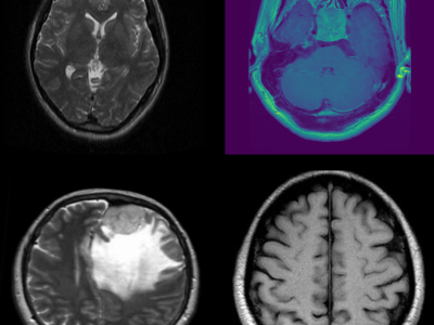
This dataset consists of MRI images of brain tumors, specifically curated for tasks such as brain tumor classification and detection. The dataset includes a variety of tumor types, including gliomas, meningiomas, and glioblastomas, enabling multi-class classification. Each MRI scan is labeled with the corresponding tumor type, providing a comprehensive resource for developing and evaluating machine learning models for medical image analysis. The data can be used to train deep learning algorithms for brain tumor detection, aiding in early diagnosis and treatment planning. It is designed to facilitate research in medical imaging and neuro-oncology, particularly in the development of automated diagnostic systems using MRI data. This dataset is suitable for researchers and practitioners working on brain cancer diagnosis, radiological imaging analysis, and deep learning-based medical image processing.
Instructions:
1. Download the Dataset:
- The dataset is provided in a single zip file. Please download the zip file from the provided link.
2. Unzip the File:
- After downloading, unzip the file. You will find two main folders:
- Training: Contains images for training your model.
- Testing: Contains images for testing and validating your model.
3. Folder Structure:
- Inside both the Training and Testing folders, there are four subfolders corresponding to different classes:
- Glioma: MRI images of patients with glioma tumors.
- Meningioma: MRI images of patients with meningioma tumors.
- Pituitary: MRI images of patients with pituitary tumors.
- No-Tumor: MRI images of patients without any tumor.
Each subfolder contains labeled images of the respective category, structured for easy use in classification tasks.
4. Usage Guidelines:
- Use the images in the Training folder for model training and the images in the Testing folder for evaluation.
- You can apply any pre-processing, augmentation, or normalization techniques suitable for MRI image analysis.
- Ensure that your model outputs correspond to the four categories: glioma, meningioma, pituitary, and no-tumor.
5. Suggested Workflow:
- Split the dataset as necessary if you need additional validation sets.
- Utilize machine learning or deep learning frameworks such as TensorFlow, PyTorch, or Keras for classification tasks.
- Implement multi-class classification techniques to distinguish between the four categories.
If you use this dataset, please be sure to cite both this work and the original datasets listed above.
The dataset is currently under embargo and will become publicly available on December 31, 2025. Until then, access is restricted to protect the integrity of the research.


great
Good luck
Best of the best
Thank you