sEMG of Larynx Functions

- Citation Author(s):
-
Johnny McNulty
(University College London, Aspire CREATe, Institute of Orthopaedics and Musculoskeletal Science London, UK)
Kylie de Jager(University College London, Aspire CREATe, Institute of Orthopaedics and Musculoskeletal Science London, UK)
Henry Lancashire(University College London, Department of Medical Physics and Biomedical Engineering London, London, UK)
James Graveston (University of Cambridge, Institute of Metabolic Science)Martin Birchall (UCL Ear Institute London, London, UK) - Submitted by:
- Johnny McNulty
- Last updated:
- DOI:
- 10.21227/6s2b-xg28
- Data Format:
 1228 views
- Categories:
- Keywords:
Abstract
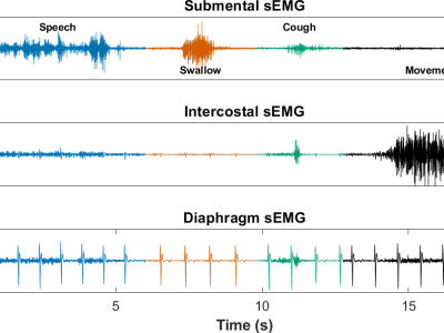
This dataset is associated with an IEEE journal submission titled: "Prediction of larynx function using multichannel surface EMG classification" by the associated authors. The dataset consists of surface electromyography (sEMG) signals recorded from 10 study participants (5 control, 5 laryngectomees), each undertaking 3 recording sessions.
During each session the following were recorded:
- 15 recordings of swallowing: 5 dry; 5 water; 5 solid (e.g. banana or biscuit).
- 3 recordings of coughing, with each recording containing 5 coughs.
- 3 recordings of speech, in which the participant read aloud 10 sentences from the Harvard sentences (IEEE, "IEEE Recommended Practice for Speech Quality Measurements, " IEEE Trans. Audio Electroacoust., vol. 17, no. 3, pp. 225-246, 1969).
- 6 recordings of movements typical of daily life (standing, sitting, reaching, twisting and walking).
Additionally, high speed video was recorded during swallowing, pneumotachometry during coughing and sound during speech.
Instructions:
1 folder for each session: "P1_S1" = Participant 1 Session 1
1 .csv file exists for each recording. Each .csv file is structured as follows:
If there are 6 columns:
- EMG-intercostal,EMG-submental,pneumotachometry,EMG-diaphragm,trigger,microphone
- mV,mV,cmH20,mV,N/A,V
If there are 7 columns:
- EMG-intercostal,EMG-submental,pneumotachometry,EMG-diaphragm,pressure,trigger,microphone
- mV,mV,cmH20,mV,V,N/A,V
Hardware setup: Two submental electrodes (EL513, 10 mm diameter, BIOPAC Systems UK) were placed on the midline, posterior to the mental protuberance, with 20 mm interelectrode distance. Three electrodes (EL503, 11 mm diameter, BIOPAC) were placed on the right 9th/10th intercostal space close to the anterior axillary line, with 35 mm interelectrode distance. The posterior two electrodes formed the intercostal recording dipole. The anterior electrode and a single electrode placed on the left 9th/10th intercostal space formed the diaphragm recording dipole. Two reference electrodes (EL503) were placed on the midline over the sternum. Two wireless EMG recorders (BIOPAC BN-EMG2 BioNomadix, 2,000 Hz sampling rate, 2,000× gain, 5 to 500 Hz bandpass filter) were placed at the waist and on the head to minimise relative cable length and motion artefacts
See README.txt for additional information


