Datasets
Standard Dataset
Normal subjects simulate hand tremor signals in Parkinson's patients
- Citation Author(s):
- Submitted by:
- Kening Gong
- Last updated:
- Mon, 07/08/2024 - 15:59
- DOI:
- 10.21227/jze1-vz79
- Data Format:
- License:
 321 Views
- Categories:
- Keywords:
Abstract
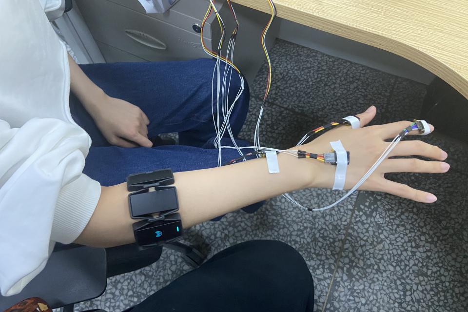
The experimental data provide hand motion signals and forearm EMG signals generated during simulated Parkinson's tremor in 10 normal subjects. The sampling frequency of the experimental equipment IMU is 100Hz, and the sampling frequency of the myoelectric armband Myo is 200Hz. The collection process of this experimental data has been approved by the Ethics Review Committee of Harbin Institute of Technology. In order to protect the privacy of the subjects, the author's consent is requested before using this data.
The motion signals are the 3-DOF acceleration, 3-DOF angular velocity and 3-DOF angle of the wrist collected by the IMU, which are given in a txt file. The EMG signal is an 8-channel surface EMG signal (μV), which is given as a csv file. All signals are given time or 16-bit unix timestamp, please calibrate the timestamp by yourself to synchronize the time of EMG signal and IMU signal.
Each subject's signal is stored in a folder, and the data in the same folder is the raw signal acquired at the same time.
The txt file saves the motion signals collected by the IMU, separated by tabs.
From left to right are: serial port, time, device number, x-axis acceleration (g), y-axis acceleration (g), z-axis acceleration (g), x-axis angular velocity (deg/s), y-axis angular velocity (deg/ s), z-axis angular velocity z(deg/s), x-axis angle (deg), y-axis angle (deg) and z-axis angular velocity (deg).
The csv file holds the sEMG signal (μV).
From left to right are: Timestamp, Channel Electrode 1, Channel Electrode 2, Channel Electrode 3, Channel Electrode 4, Channel Electrode 5, Channel Electrode 6, Channel Electrode 7 and Channel Electrode 8
Documentation
| Attachment | Size |
|---|---|
| 771 bytes |


