Datasets
Standard Dataset
Upper Limb Rehabilitation Motor Imagery EEG Signals
- Citation Author(s):
- Submitted by:
- jingfeng bi
- Last updated:
- Sun, 03/17/2024 - 05:04
- DOI:
- 10.21227/f1m3-fh49
- Data Format:
- Research Article Link:
- Links:
- License:
 695 Views
- Categories:
- Keywords:
Abstract
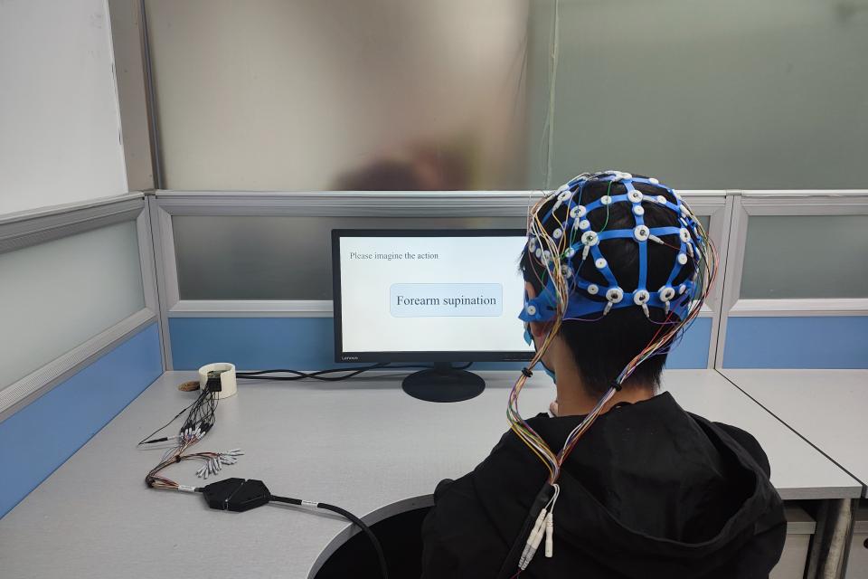
This dataset consists of electroencephalography (EEG) data from 6 participants aged between 23 and 28 years, with a mean age of 25 years. The dataset is the motor imagery EEG signals of six different rehabilitation training movements in the upper limbs. We recruited six participants aged between 23 and 28 years, with a mean age of 25 years. Three of the participants are male. Subjects sat in a comfortable chair, facing the computer monitor that displayed the trial-based paradigm and their right arm was naturally placed on the table to avoid muscle fatigue. The subjects performed six classes of motor imagination comprised of shoulder abduction, shoulder adduction, elbow flexion, elbow extension, forearm supination, forearm pronation, all about the right upper limb. Throughout the experiment, the right upper limb remained in a neutral position: the hand half open, the lower arm extended to 120 degree and in a neutral rotation.
Participants
We recruited six participants aged between 23 and 28 years, with a mean age of 25 years. Three of the participants are male.
Hardware and Software
OpenBCI CytonDaisy 16-channel Biosensing Board.
Experimental design
Subjects sat in a comfortable chair, facing the computer monitor that displayed the trial-based paradigm and their right arm was naturally placed on the table to avoid muscle fatigue. The subjects performed six classes of motor imagination comprised of shoulder abduction, shoulder adduction, elbow flexion, elbow extension, forearm supination, forearm pronation, all about the right upper limb. Throughout the experiment, the right upper limb remained in a neutral position: the hand half open, the lower arm extended to 120 degree and in a neutral rotation.
Data Records
EEG signals were recorded using 16 active Ag/AgCl electrodes via the OpenBCI CytonDaisy 16-channel Biosensing Board. An 8th order Chebyshev bandpass filter ranging from 0.01 Hz to 200 Hz was applied, and power line interference was suppressed with a notch filter at 50 Hz. The sampling frequency was set at 500 Hz, with the reference electrode placed on the left earlobe and the ground on the right earlobe, following the international standard 10-20 electrode system for EEG placement, as shown in Fig. 1. The reference electrode was placed on the left earlobe and the ground on the right earlobe. Table 1 shows the channel labels indicating the EEG electrode positions (1-16).
Data Set Description
The dataset comprises of .set files for each subject, session and run. The cues are coded as events, see Table 2 for the corresponding event codes.
Ethical issues
The study was conducted in accordance with the Declaration of Helsinki, and all subjects provided their informed consent. Ethical approval was not required for this study due to its non-invasive nature, use of anonymous data, and adherence to the Helsinki Declaration.
Documentation
| Attachment | Size |
|---|---|
| 180.48 KB |


