Datasets
Standard Dataset
Tutorial for Partial volume effect correction by Müller-Gärtner method (pvcPET)
- Citation Author(s):
-
RadekJanca
 Czech Technical University in Prague –Faculty of Electrical Engineering
 - Submitted by:
- Radek Janca
- Last updated:
- Thu, 06/15/2023 - 05:43
- DOI:
- 10.21227/z3e0-bh73
- Data Format:
- License:
 547 Views
- Categories:
- Keywords:
Abstract
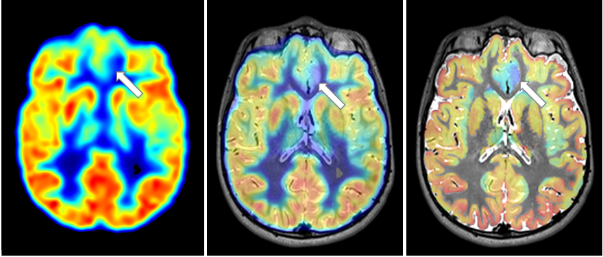
Neuroimaging methods play an important role in presurgical examinations and localization of epileptogenic lesion. Magnetic resonance imaging (MRI) is a neuroimaging technique that is essential to detect structurally abnormal tissue and thus delineate the epileptogenic lesion. Magnetic resonance imaging (MRI) provides structural data and can reveal underlying epileptogenic lesions (T1, T2, FLAIR). Epilepsy surgery candidates are often divided based on the detailed magnetic resonance findings into lesional (underlying structural pathology visible on MRI) and non-lesional cases (with normal brain MRI). Especially non-lesional cases (up to 50% of cases) represent the most challenging group of patients with a complicated epilepsy surgery. Positron emission tomography of 2-deoxy-2-[18F]fluoro-D-glucose radiotracer (FDG-PET) is a functional neuroimaging method that can also significantly contribute to the localization of the epileptogenic lesion. FDG-PET image visualizes the tissue metabolic activity associated with the consumption of glucose and perfusion. The tissue of epileptogenic lesion often shows decreased metabolic activity (hypometabolism) and in the FDG-PET image it can be recognized as a region with hypointense signal.
However, the hypometabolic lesions can be very subtle and their identification in the MRI and PET image might be still difficult and easy to miss. One of the important effects that limits the use of FDG-PET for precise localization of the subtle hypometabolic lesions is the low effective spatial resolution which leads to blurring of the PET image. In comparison, effective resolution of modern 3T MRI is better than 1 mm, but PET effective resolution varies from 3 to 8 mm depends on PET scanner generation and mainly physical limitations. PET scanner measures glucose metabolism indirectly using beta+ decay of radiotracer in FDG. Positrons are emited from a point source of FDG accumulation. It scatters through matter (»1 mm for) losing energy and annihilates with an electron resulting in two detectable 511 keV photons that are emitted in nearly opposite directions (180±0.5°). This, together with other physical effects, causes that the point source is imaged as the point spread function (PSF) and results in naturally blurred PET images with effective. The blurring grows from the centre due to the increased probability of false LOR detection in neighbouring detecting crystals absorbing 511keV gamma photons. The signals emitted from neighbouring tissue mix together which results in partial volume effect (PVE). For example, naturally metabolic grey matter is mixed with hypometabolic white matter and other tissue, that leads to an underestimation of radiotracer activity in the cortex and blurring of tissue edges. This effect can lead to spurious hypometabolic regions, resulting in an increased amount of false-positive hypometabolic regions, and vice versa.
Partial volume effect can be compensated using partial volume correction (PVC) which can additionally increase the effective spatial resolution of the PET image. Various methods for partial volume correction have been proposed [1]. One of the important inputs for partial volume correction methods is the full width at half maximum (FWHM) of the scanner point spread function (PSF), which defines how much point source is blurred.
PVC will be performed with Müller-Gärtner method [2, 3] using implementation from publicly available PETPVC toolbox. Müller-Gärtner method is one of the post-reconstruction methods for PVC, which performs correction of radiotracer activity for each voxel back to the grey matter tissue segment. In summary, the technique refocuses PET activity to grey matter well defined in MRI.
[1] Erlandsson, K., Buvat, I., Pretorius, P. H., Thomas, B. A., & Hutton, B. F. (2012). A review of partial volume correction techniques for emission tomography and their applications in neurology, cardiology and oncology. Physics in Medicine & Biology, 57(21), R119.
[2] Müller-Gärtner, H. W., Links, J. M., Prince, J. L., Bryan, R. N., McVeigh, E., Leal, J. P., ... & Frost, J. J. (1992). Measurement of radiotracer concentration in brain gray matter using positron emission tomography: MRI-based correction for partial volume effects. Journal of Cerebral Blood Flow & Metabolism, 12(4), 571-583.
[3] Thomas, B. A., Cuplov, V., Bousse, A., Mendes, A., Thielemans, K., Hutton, B. F., & Erlandsson, K. (2016). PETPVC: a toolbox for performing partial volume correction techniques in positron emission tomography. Physics in Medicine & Biology, 61(22), 7975.
The workflow of processing is described in MATLAB live script ILAE_pvcPET_v?.mlx
More from this Author

Dataset Files
- Starting dataset and function package ILAE_AMIE_SuSIE_2023.zip (80.64 MB)
- Processed files ILAE_AMIE_SuSIE_2023 - processed.zip (770.40 MB)
- Basic tutorial of PET-MRI registration in 3D Slicer and DICOM data sample Multimodal registration by Slicer.zip (114.56 MB)
Documentation


