Mouse brain lightfield-confocal stack dataset
- Citation Author(s):
-
Josue Page
 (Computational Imaging and Inverse Problems Group, Technical University of Munich)
Federico Saltarin
(Theodor Kocher Institute, University of Bern, Switzerland)
Yury Belyaev
(Microscopy Imaging Center, University of Bern, Switzerland)
Ruth Lyck
(Theodor Kocher Institute, University of Bern, Switzerland)
Tobias Lasser
(Computational Imaging and Inverse Problems Group, Technical University of Munich)
Paolo Favaro
(Computer Vision Group, University of Bern, Switzerland)
(Computational Imaging and Inverse Problems Group, Technical University of Munich)
Federico Saltarin
(Theodor Kocher Institute, University of Bern, Switzerland)
Yury Belyaev
(Microscopy Imaging Center, University of Bern, Switzerland)
Ruth Lyck
(Theodor Kocher Institute, University of Bern, Switzerland)
Tobias Lasser
(Computational Imaging and Inverse Problems Group, Technical University of Munich)
Paolo Favaro
(Computer Vision Group, University of Bern, Switzerland)
- Submitted by:
- Josue Page
- Last updated:
- DOI:
- 10.21227/eagc-ta82
- Data Format:
- Research Article Link:
- Links:
 646 views
- Categories:
- Keywords:
Abstract
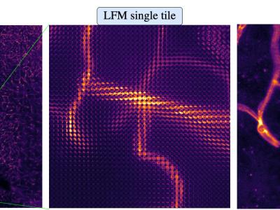
Dataset of fluorescent mice brain vessels Confocal 3D volumes aligned to Light-Field images.
Confocal:
- Single volume dimension: 1287x1287x64.
- Number of samples: 362
- Voxel size: 0.086x0.086x0.9 um.
- Objective: 40x/1.3 Oil.
- Stain: tomato lectin (DyLight594 conjugated, DL-1177, Vector Laboratories).
LightField:
- Image dimensions 1287x1287.
- PixelSize: 3.45 um.
- Pixels per lenslet: 33x33.
- Lenslet Pitch: 112 um.
- MLA2Sensor distance: 2500um.
- Tube-lens focal length: 165mm.
- Objective: 40x/0.9 Air.
H5 containing:
- volData: confocal volumes 1287x1287x64 voxels.
- LFData: LF 4D tensor 33x33x39x39 (Angular coord x, angular coord y, spatial coord x, spatial coord y).
- gridCoords: image grid positions.
Instructions:
Rename the file extension from hdf5 to .h5