Datasets used in "3-D quantification of filopodia in motile cancer cells" IEEE Transactions on Medical Imaging 38(3), 862-872 (2019)
- Citation Author(s):
-
Carlos Castilla (Center for Applied Medical Research, University of Navarra, Pamplona, Spain)Martin Maska (Centre for Biomedical Image Analysis Faculty of Informatics, Masaryk University, Brno, Czech Republic)Dmitry Sorokin (Centre for Biomedical Image Analysis Faculty of Informatics, Masaryk University, Brno, Czech Republic)Erik Meijering (Departments of Medical Informatics and Radiology, Erasmus University Medical Center Rotterdam, The Netherlands)
- Submitted by:
- Carlos Ortiz De Solorzano
- Last updated:
- DOI:
- 10.21227/H26W9K
- Research Article Link:
- Links:
 702 views
- Categories:
Abstract
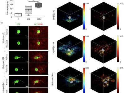
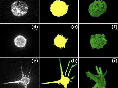
These datasets were used to produce the results of the following TMI paper: "3D Quantification of Filopodia in Motile Cancer Cells", Castilla C., et al. (2019). IEEE Transactions on Medical Imaging 38(3):862,872.
3D+t image real and synthetic sequences of cells of the A549 lung adenocarcinoma cancer cell line, displaying three different phenotypes of CRMP-2, a protein involved in the assembly and disassembly of actin filaments. All real videos were acquired on aUltraviewERS (Perkin Elmer, Inc., Waltham, MA, USA), spinning disk confocal microscope, using the 488 nm line of an Ar/Kr laser to image the sample through a Plan-Apochromatic 63x 1.20 NA water immersion objective lens (Carl Zeiss, AG., Wetzlar, Germany). The videos contained one cell, imaged every two minutes duringone hour. The original image voxel size of 0.126x0.126x1.0 µm was resampled in the axial direction using cubic spline interpolation to obtain isotropic image data with a voxel size of 0.12x0.126x0.126 µm. The synthetic videos were produced using our recent cell simulator [1], to reproduce as closely as possible the image properties and cell phenotypes of the real videos.
Along with the datasets, we provide AVI videos displaying the results of maximum intensity projections (MIP) of the segmentation of the cells using two segmenation methods, one based on the minimization of the Chan-Vese model (CVS) and a 3D Convolutional Neural Network (CNN)
The file names encode the following information:
CRMP2 phenotype and video type:
- WTR: Wild type, real video
- OER: Over expressing, real video
- PDR: Phospho-defective, real video
- WTS: Wild type, synthetic video
- OES: Over expressing, synthetic video
- PDS: Phospho-defective, synthetic video
Segmentation method:
- CVS: Minimization of the Chan-Vese models
- CNN: Convolutional Neural Network
Video number:
- 01
- 02
- 03
[1] D. V. Sorokin, I. Peterlík, V. Ulman, D. Svoboda and M. Maška, “Model-based generation of synthetic 3D time-lapse sequences of motile cells with growing filopodia,” In IEEE International Symposium on Biomedical Imaging, pp. 822–826, 2017
Instructions:
Each zip file contains a set of 3D image frames.
Dataset Files
- CRMP2 Wild Type Real video sequence 1 (Size: 42.3 MB)
- CRMP2 Wild Type Real video sequence 2 (Size: 43.06 MB)
- CRMP2 Wild Type Real video sequence 3 (Size: 41.73 MB)
- CRMP2 Overexpressing Real video sequence 1 (Size: 129.77 MB)
- CRMP2 Overexpressing Real video sequence 2 (Size: 126.89 MB)
- CRMP2 Overexpressing Real video sequence 3 (Size: 131.64 MB)
- CRMP2 Phosphodefective Real video sequence 1 (Size: 162.96 MB)
- CRMP2 Phosphodefective Real video sequence 2 (Size: 339.28 MB)
- CRMP2 Phosphodefective Real video sequence 3 (Size: 116.21 MB)
- CRMP2 Wild Type Synthetic video sequence 1 (Size: 129.71 MB)
- CRMP2 Wild Type Synthetic video sequence 2 (Size: 98.56 MB)
- CRMP2 Wild Type Synthetic video sequence 3 (Size: 132.48 MB)
- CRMP2 Overexpressing Synthetic video sequence 1 (Size: 171.76 MB)
- CRMP2 Overexpressing Synthetic video sequence 2 (Size: 176.45 MB)
- CRMP2 Overexpressing Synthetic video sequence 3 (Size: 216.18 MB)
- CRMP2 Phosphodefective Synthetic video sequence 1 (Size: 905.23 MB)
- CRMP2 Phosphodefective Synthetic video sequence 2 (Size: 878.52 MB)
- CRMP2 Phosphodefective Synthetic video sequence 3 (Size: 825.03 MB)
- MIP Video results CRMP2 Wild Type Real video sequence 1 CVS method (Size: 219.35 KB)
- MIP Video results CRMP2 Wild Type Real video sequence 2 CVS method (Size: 219.35 KB)
- MIP Video results CRMP2 Wild Type Real video sequence 3 CVS method (Size: 219.97 KB)
- MIP Video results CRMP2 Overexpressing Real video sequence 1 CVS method (Size: 279.79 KB)
- MIP Video results CRMP2 Overexpressing Real video sequence 2 CVS method (Size: 293.89 KB)
- MIP Video results CRMP2 Overexpressing Real video sequence 3 CVS method (Size: 328.08 KB)
- MIP Video results CRMP2 Phosphodefective Real video sequence 1 CVS method (Size: 547.44 KB)
- MIP Video results CRMP2 Phosphodefective Real video sequence 2 CVS method (Size: 788.61 KB)
- MIP Video results CRMP2 Phosphodefective Real video sequence 3 CVS method (Size: 275.94 KB)
- MIP Video results CRMP2 Wild Type Synthetic video sequence 1 CVS method (Size: 190.48 KB)
- MIP Video results CRMP2 Wild Type Synthetic video sequence 2 CVS method (Size: 190.79 KB)
- MIP WTS 03 CVS.avi (Size: 201.59 KB)
- MIP Video results CRMP2 Overexpressing Synthetic video sequence 1 CVS method (Size: 192.38 KB)
- MIP Video results CRMP2 Overexpressing Synthetic video sequence 2 CVS method (Size: 195.41 KB)
- MIP Video results CRMP2 Overexpressing Synthetic video sequence 3 CVS method (Size: 217.11 KB)
- MIP Video results CRMP2 Phosphodefective Synthetic video sequence 1 CVS method (Size: 476.58 KB)
- MIP Video results CRMP2 Phosphodefective Synthetic video sequence 2 CVS method (Size: 360.98 KB)
- MIP Video results CRMP2 Phosphodefective Synthetic video sequence 3 CVS method (Size: 412.14 KB)
- MIP Video results CRMP2 Wild Type Real video sequence 1 CNN method (Size: 188.2 KB)
- MIP Video results CRMP2 Wild Type Real video sequence 2 CNN method (Size: 192.66 KB)
- MIP Video results CRMP2 Wild Type Real video sequence 3 CNN method (Size: 190.88 KB)
- MIP Video results CRMP2 Overexpressing Real video sequence 1 CNN method (Size: 267.3 KB)
- MIP Video results CRMP2 Overexpressing Real video sequence 2 CNN method (Size: 277.02 KB)
- MIP Video results CRMP2 Overexpressing Real video sequence 3 CNN method (Size: 290.6 KB)
- MIP Video results CRMP2 Phosphodefective Real video sequence 1 CNN method (Size: 586.09 KB)
- MIP Video results CRMP2 Phosphodefective Real video sequence 2 CNN method (Size: 943.6 KB)
- MIP Video results CRMP2 Phosphodefective Real video sequence 3 CNN method (Size: 326.76 KB)
- MIP Video results CRMP2 Wild Type Synthetic video sequence 1 CNN method (Size: 185.96 KB)
- MIP Video results CRMP2 Wild Type Synthetic video sequence 2 CNN method (Size: 181.43 KB)
- MIP Video results CRMP2 Wild Type Synthetic video sequence 3 CNN method (Size: 200.52 KB)
- MIP Video results CRMP2 Overexpressing Synthetic video sequence 1 CNN method (Size: 218.59 KB)
- MIP OES 02 CNN.avi (Size: 213.09 KB)
- MIP OES 03 CNN.avi (Size: 247.07 KB)
- MIP Video results CRMP2 Phosphodefective Synthetic video sequence 1 CNN method (Size: 609.58 KB)
- MIP Video results CRMP2 Phosphodefective Synthetic video sequence 2 CNN method (Size: 482.55 KB)
- MIP Video results CRMP2 Phosphodefective Synthetic video sequence 3 CNN method (Size: 476.08 KB)


