CT AND MRI FUSION
- Citation Author(s):
-
yuping huang
 (hongqing Key Laboratory of Image Cognition, Chongqing University of Posts and Telecommunications)
- Submitted by:
- yuping huang
- Last updated:
- DOI:
- 10.21227/24x1-a288
- Data Format:
- Research Article Link:
 522 views
- Categories:
- Keywords:
Abstract
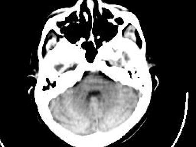
CT (roentgen-ray computed tomography) A beam of x-rays is shot straight through the brain. As it comes out the other side, the beam is blunted slightly because it has hit dense living tissues on the way through. Blunting or "attenuation" of the x-ray comes from the density of the tissue encountered along the way. Very dense tissue like bone blocks lots of x-rays; grey matter blocks some and fluid even less. X-ray detectors positioned around the circumference of the scanner collect attenuation readings from multiple angles. A computerized algorithm reconstructs an image of each slice
Instructions:
MRI (magnetic resonance imaging) When protons (here brain protons) are placed in a magnetic field, they become capable of receiving and then transmitting electromagnetic energy. The strength of the transmitted energy is proportional to the number of protons in the tissue. Signal strength is modified by properties of each proton's microenvironment, such as its mobility and the local homogeneity of the magnetic field. MR signal can be "weighted" to accentuate some properties and not others.


verry god
good