Datasets
Standard Dataset
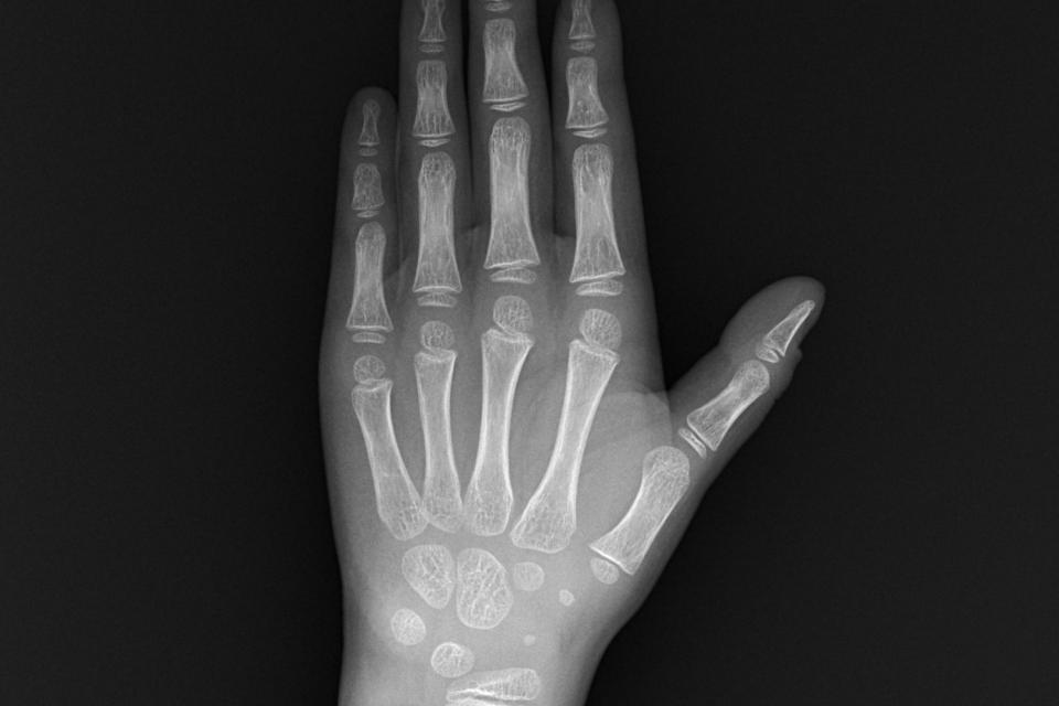
wrist X-ray
- Citation Author(s):
- Submitted by:
- fang kai
- Last updated:
- Mon, 07/08/2024 - 15:59
- DOI:
- 10.21227/75h6-8a71
- Data Format:
- License:
 203 Views
- Categories:
- Keywords:
Abstract
The X-ray image of wrist bone is collected by professional sensing equipment. The radiation of the device is small enough to ensure the health of the subject. In order to ensure the standardization of X-ray images, remove all the handpieces of the left hand during the shooting process. The five fingers of the left hand should be naturally extended, and the palm should be kept downward. In addition, when shooting, the middle finger and forearm must be as straight as possible. The captured images are in DICOM format, with a size of 1626 × 2032 pixels. We convert the image format to jpg style, and desensitize personal information such as name and ID number in the dataset.
In the dataset file, images are the x-ray storage folder, Annotations are the xml files that store the location information of each reference bone in each picture, and the label folder is the storage location of the corresponding labels of each reference bone in each x-ray. You can use this data set to perform work related to reference bone cutting.
Documentation
| Attachment | Size |
|---|---|
| 53.14 KB |


Comments
Thank you for sharing this dataset!
Can you please describe the formatting for the label .txt files in this dataset?
Each of the label .txt files has 14 rows and each row has five entries, where the first one is always an integer, and the remaining ones are double precision fractions.
What do the integers and the numbers following them represent?
How can the provided labels be related to the corresponding .jpg images?
Thank you!