Leucorrhea Microscopy Dataset
- Citation Author(s):
- Submitted by:
- Xiaohui Du
- Last updated:
- DOI:
- 10.21227/8rcr-cr03
 450 views
- Categories:
- Keywords:
Abstract
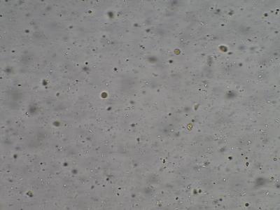
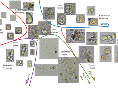
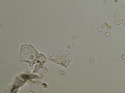
Leucorrhea microscopic data set is a set of leucorrhea microscopic images, which is used in object detection task. The datasets are collected from the Sixth People’s Hospital of Chengdu (Sichuan Province, China). The samples were went flow diluted, stirred and placed, and imaged with a microscopic imaging system. The clearest 3 images were collected for each view of each sample with Tenengrad definition algorithm. The dataset we collected includes 1552 groups of views with 4656 jpg images. The Resolution of images are 1200×1920. There are 6 categories, RBCs, WBCs, Molds, Epithelial Cells, Pyocytes and Trichomonads. Each image folder is a sample collected with different views, namely H-%1-%2, and %1 represents for the view id, and %2 is the image id.
Instructions:
The clearest 3 images represent the image captured from object distance. For the object detection of different object distance images in a field of vision, we define it as super depth of field (SDoF) detection.
The annotation is described in the CSV file, the format is as followed:
view pic type x y width height isValid label
1 1 Epi 10 20 400 600 1 1
The column view is the view id, the pic represents the image id. Type is the category name of a cell. X\y\width\height are the bounding box of the target. isValid is the object is valid or not. And the label is the count id of the current image.


