Left Atrium 2018
- Citation Author(s):
-
Hengfan Li (Jiangsu Collaborative Innovation Center for Atmospheric Environment and Equipment Technology, Nanjing University of Information Science and TechnologyJiangsu Collaborative Innovation Center for Atmospheric Environment and Equipment Technology, Nanjing Uni)
- Submitted by:
- Hengfan Li
- Last updated:
- DOI:
- 10.21227/5y8c-wf96
- Data Format:
 147 views
- Categories:
- Keywords:
Abstract
The Left Atrium2018 dataset was used in the 2018 Left Atrium Segmentation Challenge and has the following characteristics:
Data Content
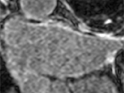
Image Type: It consists of 154 three-dimensional gadolinium-enhanced magnetic resonance imaging (LGE-MRI) images, which is currently the largest cardiac LGE-MRI dataset in the world.
Label Information: It contains the relevant labels of the left atrium segmented by three medical experts.
Application Fields
It is mainly applied in the field of medical image segmentation, especially in the segmentation and visualization research of the left atrium, which is of great significance for the clinical diagnosis and treatment of diseases such as atrial fibrillation.
Public Access
The dataset can be obtained at [https://www.cardiacatlas.org/atria seg2018-challenge/](https://www.cardiacatlas.org/atria seg2018-challenge/).
The following is the general usage method:
Data Preprocessing
Format Conversion: According to the requirements of the used deep learning framework and model, convert the original data format, such as converting the image data into a tensor format suitable for model input.
Normalization: Normalize the image data, scaling the pixel values to a certain range, such as [0, 1] or [-1, 1], to improve the training effect and convergence speed of the model.
Data Augmentation: To increase the diversity of data and the generalization ability of the model, data augmentation operations can be carried out, such as random flipping, rotation, scaling, cropping, etc. For example, in PyTorch, the torchvision.transforms module can be used to achieve data augmentation.
Model Selection and Training
Select a Suitable Model: According to the task requirements and the characteristics of the dataset, select a suitable deep learning model, such as U-Net, V-Net, 3D-UNet and other commonly used models for medical image segmentation.
Divide the Dataset: Divide the dataset into a training set, a validation set, and a test set. Generally, it can be divided according to a ratio of 7:1:2 or 8:1:1.
Model Training: Use the training set to train the model. During the training process, adjust the parameters of the model and the optimization algorithm to make the model gradually fit the data. For example, in PyTorch, the torch.nn module can be used to build the model, and the torch.optim module can be used to select optimization algorithms, such as Stochastic Gradient Descent (SGD), Adagrad, Adadelta, Adam, etc., and then use the training data to iteratively train the model.
Model Evaluation and Optimization: Use the validation set to evaluate the trained model, and adjust the hyperparameters of the model according to the evaluation results, such as learning rate, number of layers, convolutional kernel size, etc., to improve the performance of the model. Commonly used evaluation metrics can be used, such as Dice coefficient, Jaccard coefficient, Average Surface Distance (ASD), 95% Hausdorff Distance (95HD), etc.
Model Testing: Use the test set to test the finally optimized model to evaluate the performance and generalization ability of the model on unknown data.
Result Analysis and Application
Visualize the Segmentation Results: Visualize the segmentation results of the model, compare them with the real labels, and intuitively observe the segmentation effect of the model, which helps to analyze the advantages and disadvantages of the model and possible problems.
Clinical Application: Apply the trained model to actual clinical diagnosis and treatment, assist doctors in the segmentation and analysis of the left atrium, and improve the accuracy and efficiency of diagnosis.
Model Improvement and Innovation: According to the experimental results and analysis, improve and innovate the model, such as proposing new network structures, loss functions, training methods, etc., to further improve the performance and effect of the model.
Instructions:
The Left Atrium2018 dataset was used in the 2018 Left Atrium Segmentation Challenge and has the following characteristics:
Data Content
Image Type: It consists of 154 three-dimensional gadolinium-enhanced magnetic resonance imaging (LGE-MRI) images, which is currently the largest cardiac LGE-MRI dataset in the world.
Label Information: It contains the relevant labels of the left atrium segmented by three medical experts.
Application Fields
It is mainly applied in the field of medical image segmentation, especially in the segmentation and visualization research of the left atrium, which is of great significance for the clinical diagnosis and treatment of diseases such as atrial fibrillation.
Public Access
The dataset can be obtained at [https://www.cardiacatlas.org/atria seg2018-challenge/](https://www.cardiacatlas.org/atria seg2018-challenge/).
The following is the general usage method:
Data Preprocessing
Format Conversion: According to the requirements of the used deep learning framework and model, convert the original data format, such as converting the image data into a tensor format suitable for model input.
Normalization: Normalize the image data, scaling the pixel values to a certain range, such as [0, 1] or [-1, 1], to improve the training effect and convergence speed of the model.
Data Augmentation: To increase the diversity of data and the generalization ability of the model, data augmentation operations can be carried out, such as random flipping, rotation, scaling, cropping, etc. For example, in PyTorch, the torchvision.transforms module can be used to achieve data augmentation.
Model Selection and Training
Select a Suitable Model: According to the task requirements and the characteristics of the dataset, select a suitable deep learning model, such as U-Net, V-Net, 3D-UNet and other commonly used models for medical image segmentation.
Divide the Dataset: Divide the dataset into a training set, a validation set, and a test set. Generally, it can be divided according to a ratio of 7:1:2 or 8:1:1.
Model Training: Use the training set to train the model. During the training process, adjust the parameters of the model and the optimization algorithm to make the model gradually fit the data. For example, in PyTorch, the torch.nn module can be used to build the model, and the torch.optim module can be used to select optimization algorithms, such as Stochastic Gradient Descent (SGD), Adagrad, Adadelta, Adam, etc., and then use the training data to iteratively train the model.
Model Evaluation and Optimization: Use the validation set to evaluate the trained model, and adjust the hyperparameters of the model according to the evaluation results, such as learning rate, number of layers, convolutional kernel size, etc., to improve the performance of the model. Commonly used evaluation metrics can be used, such as Dice coefficient, Jaccard coefficient, Average Surface Distance (ASD), 95% Hausdorff Distance (95HD), etc.
Model Testing: Use the test set to test the finally optimized model to evaluate the performance and generalization ability of the model on unknown data.
Result Analysis and Application
Visualize the Segmentation Results: Visualize the segmentation results of the model, compare them with the real labels, and intuitively observe the segmentation effect of the model, which helps to analyze the advantages and disadvantages of the model and possible problems.
Clinical Application: Apply the trained model to actual clinical diagnosis and treatment, assist doctors in the segmentation and analysis of the left atrium, and improve the accuracy and efficiency of diagnosis.
Model Improvement and Innovation: According to the experimental results and analysis, improve and innovate the model, such as proposing new network structures, loss functions, training methods, etc., to further improve the performance and effect of the model.